Complete Guide to Testing, Treatment, and Prevention of Internal Parasites (Protozoans and Roundworms) in Reptiles
Explains the types and symptoms of common internal parasites in reptiles (protozoans, nematodes, roundworms, and tapeworms). Detailed coverage of fecal testing methods, types of antiparasitic drugs and precautions, differences between WC and domestic CB specimens, and the pros and cons of preventive treatment.
Key Takeaways
Explains the types and symptoms of common internal parasites in reptiles (protozoans, nematodes, roundworms, and tapeworms). Detailed coverage of fecal testing methods, types of antiparasitic drugs and precautions, differences between WC and domestic CB specimens, and the pros and cons of preventive treatment.
Related Species
Reptile Parasite Problems: Why It Matters
Reptiles, particularly WC (wild-caught) specimens, commonly harbor internal parasites. In Japan, internal parasites in reptiles are often overlooked, but continuing to keep infested animals without treatment can lead to deteriorated health, immunosuppression, chronic anorexia, and in the worst cases, death.
Even domestic CB (captive-bred) or well-established captive-bred specimens are not completely parasite-free. Particularly when fed live food insects such as crickets, these insects can act as intermediate hosts and transmit parasites. In multi-specimen enclosures, parasite transmission via feces occurs more readily.
Internal parasites in reptiles are broadly divided into two categories: "protozoans" and "helminths (parasitic worms)." Because symptoms, testing methods, and antiparasitic medications differ for each, identifying what parasite is present is the first step in treatment.
Protozoan Infections: Cryptosporidium, Coccidium, and Amoeba
Protozoans are single-celled microorganisms, and the following are representative parasites of reptiles.
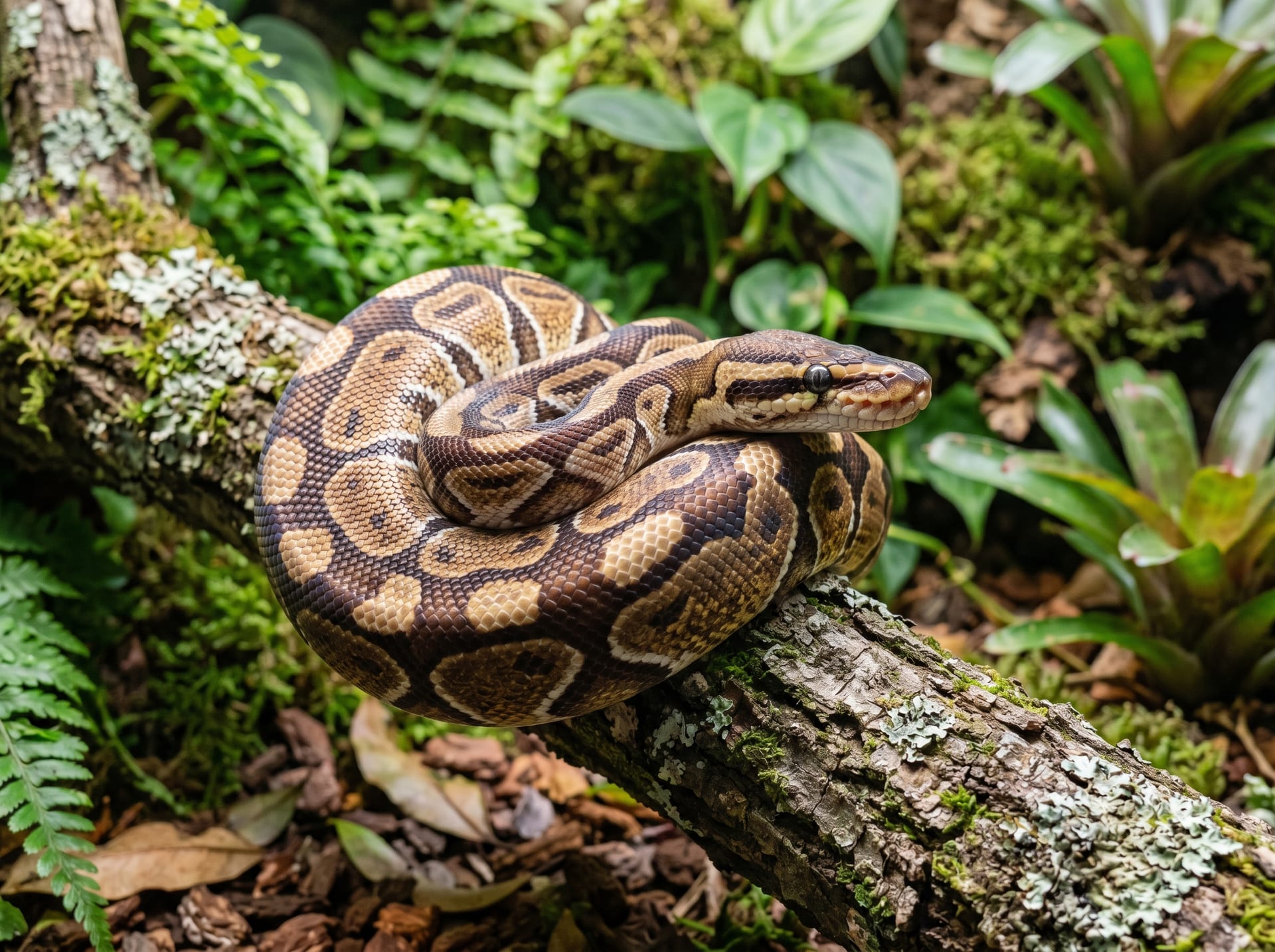
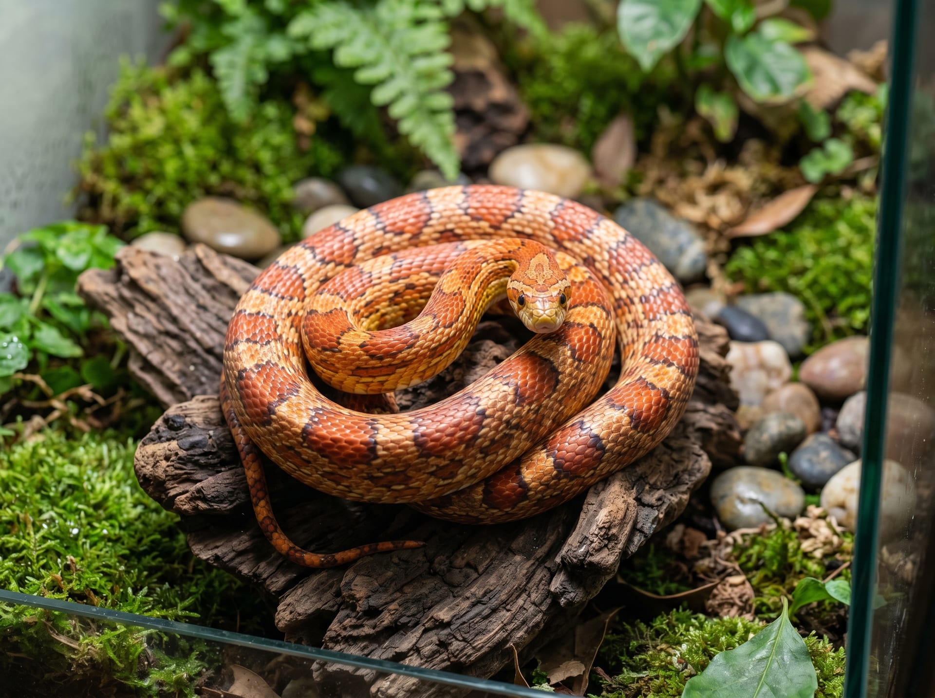
Cryptosporidium is one of the most serious protozoan parasites in reptile medicine. It commonly affects ball pythons and corn snakes, and when infected, it colonizes the gastrointestinal mucosa and impairs digestion and absorption. The characteristic symptom is mid-body swelling, accompanied by anorexia, weight loss, and repeated vomiting. Unfortunately, no definitive treatment is currently established, and infected individuals often become lifelong carriers. It has extremely high transmission potential to other animals and spreads easily through cohabitation, shared enclosure items, and inadequate fecal management.
Coccidium (genus Eimeria, Isospora, etc.) parasitizes the digestive tract and causes diarrhea, bloody feces, and lethargy in juveniles or immunocompromised individuals. Because oocysts (spoulated eggs) are shed in feces, environmental contamination rapidly progresses in warm, humid conditions. Trimethoprim-sulfadiazine and ponazuril are commonly used for treatment (always follow veterinary instructions).
Amoeba (amoebic dysentery-related organisms) have been reported in tortoises and lizards and can cause enteritis and necrotic lesions.
Helminth Infections: Characteristics of Roundworms, Tapeworms, and Hookworms
Helminths (parasitic worms) are multicellular parasites of visible size that parasitize the digestive tract and tissues.
Roundworms (nematodes: Ascarididae) are widely seen in snakes and lizards and can grow in the digestive tract, causing nutritional competition, intestinal obstruction, and bowel perforation. Early infection is often asymptomatic, and discovery often first occurs through fecal examination.
Tapeworms (Taenia species) are transmitted via live prey (mice, crickets, mealworms) as intermediate hosts and grow in the intestinal tract. Cases are sometimes detected when white rice-grain-like segments appear in the feces.
Hookworms and whipworms (Ancylostoma species and Trichuris species) burrow into the intestinal mucosa and feed on blood. Chronic infections result in anemia, hypoproteinemia, and edema.
Fenbendazole and ivermectin-based medications are commonly used against helminths in general, but dosage, dosing intervals, and contraindications (ivermectin use in snakes requires special caution) vary by species, so always consult with a reptile-experienced veterinarian.
Testing Process: Fecal Examination and Specialist Consultation
Fecal examination (flotation and smear techniques) is fundamental for confirming internal parasite diagnosis. Fresh feces (ideally within 2-4 hours of defecation) should be placed in a small sealed container and brought to a veterinary clinic that offers reptile diagnostics.
The examination uses microscopy to confirm the presence or absence of eggs, oocysts, and cysts. However, detection rates may be low in a single test, so repeat testing is recommended if symptoms persist. Cryptosporidium is difficult to detect by routine flotation and may require specialized testing such as acid-fast staining, ELISA, or PCR.
When acquiring new specimens, especially WC animals, fecal testing is strongly recommended within 1-2 weeks of acquisition, even if asymptomatic. This "introduction screening" is one of the most effective ways to prevent transmission to established animals.
Antiparasitic Treatment in Practice and Precautions
Antiparasitic medications are prescribed by a veterinarian after considering the animal's species, weight, parasite type, and health status. Owners should avoid self-administering over-the-counter animal medications, as this risks toxicity from overdosing and development of resistance from inappropriate treatment.
Pay attention to the following points during treatment.
- Thorough environmental cleaning: Even after parasites are killed by medication, reinfection can occur from eggs or cysts remaining in the enclosure, substrate, and water bowl. Disinfection with ammonia-based cleaners or high-temperature steam is effective (alcohol and chlorine-based disinfectants may be ineffective against some protozoan eggs).
- Post-treatment fecal retest: Perform fecal examination again 2-4 weeks after treatment completion to confirm parasite elimination.
- Isolation management: In multi-specimen setups, infected individuals must be completely separated from other animals until treatment is complete and negativity is confirmed.
- Appetite and hydration monitoring: Temporary appetite suppression and soft feces may occur as side effects of antiparasitic medication. Return for follow-up if lethargy or vomiting persists.
Daily Management for Prevention
Prevention of internal parasites comes down to maintaining a clean husbandry environment and blocking infection sources.
Live food management: Obtain insects such as crickets and dubia roaches from reliable suppliers with proper hygiene standards and avoid excessive stockpiling. Feeding wild-collected insects carries extremely high parasite transmission risk and should be avoided.
Prompt fecal removal: Remove feces as soon as possible after defecation and perform regular complete substrate changes. Oocysts such as coccidium can survive in the environment for weeks to months.
Limiting multi-specimen housing: Housing multiple animals in the same enclosure greatly increases infection risk. Ideally, each individual should have a separate enclosure.
Regular health checks: Annual or bi-annual fecal examinations can detect asymptomatic infections even without symptoms. Since reptiles tend to hide symptoms, appearing healthy does not ensure safety.
BreederDirect handles specimens with documented parasite screening and treatment history. For any questions about an individual's health status, please contact the seller directly or consult with a reptile specialist veterinarian.