Dog Patellar Luxation: Symptoms, Grading, Surgery, and Management Guide
A comprehensive guide to patellar luxation (patella) in small breeds, covering symptoms, severity grades (1–4), surgical necessity assessment, post-operative rehabilitation methods, and preventive muscle-strengthening exercises. Essential information for owners of Chihuahuas, Pomeranians, and Toy Poodles.
Key Takeaways
A comprehensive guide to patellar luxation (patella) in small breeds, covering symptoms, severity grades (1–4), surgical necessity assessment, post-operative rehabilitation methods, and preventive muscle-strengthening exercises. Essential information for owners of Chihuahuas, Pomeranians, and Toy Poodles.
Related Species
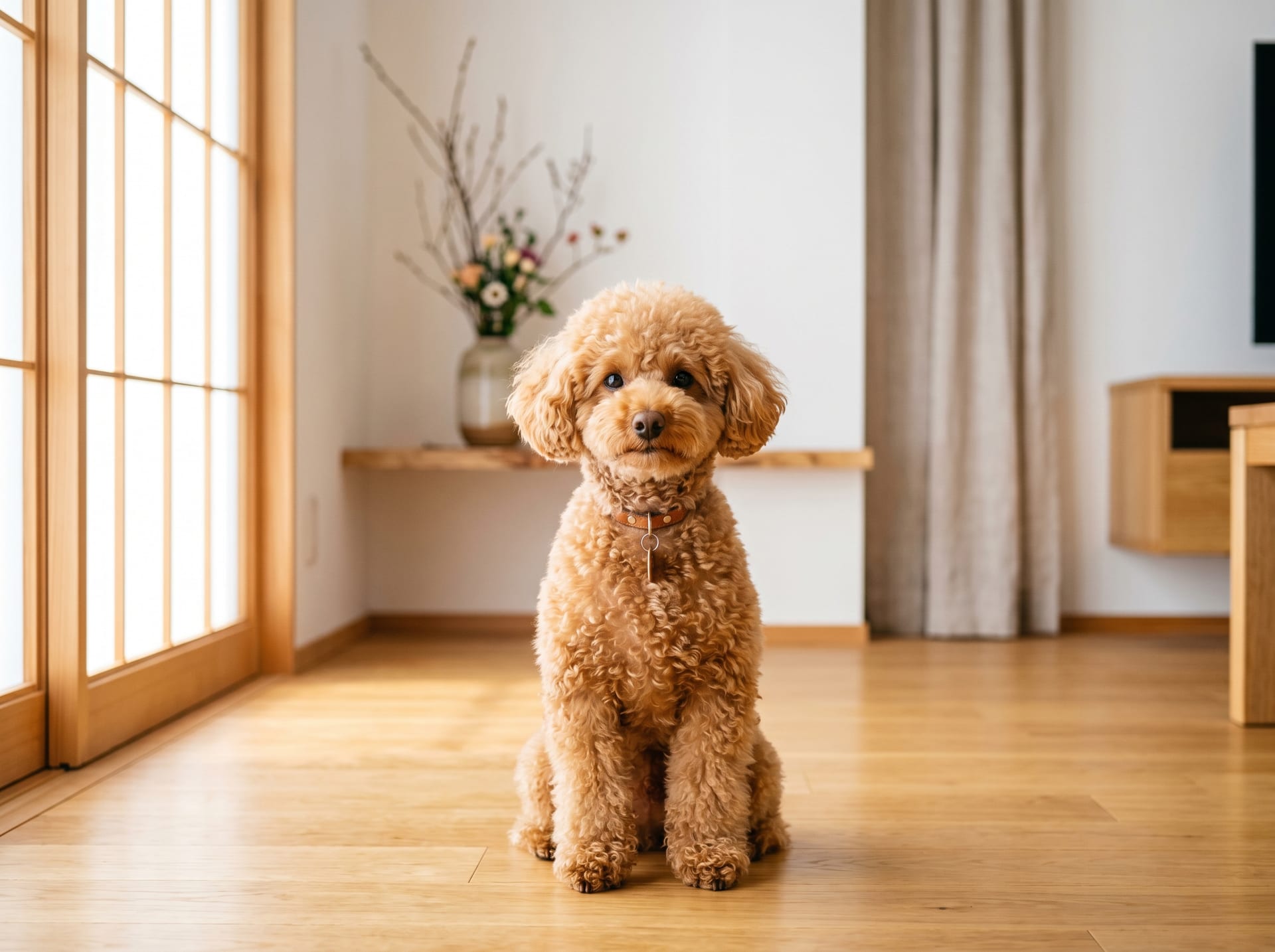
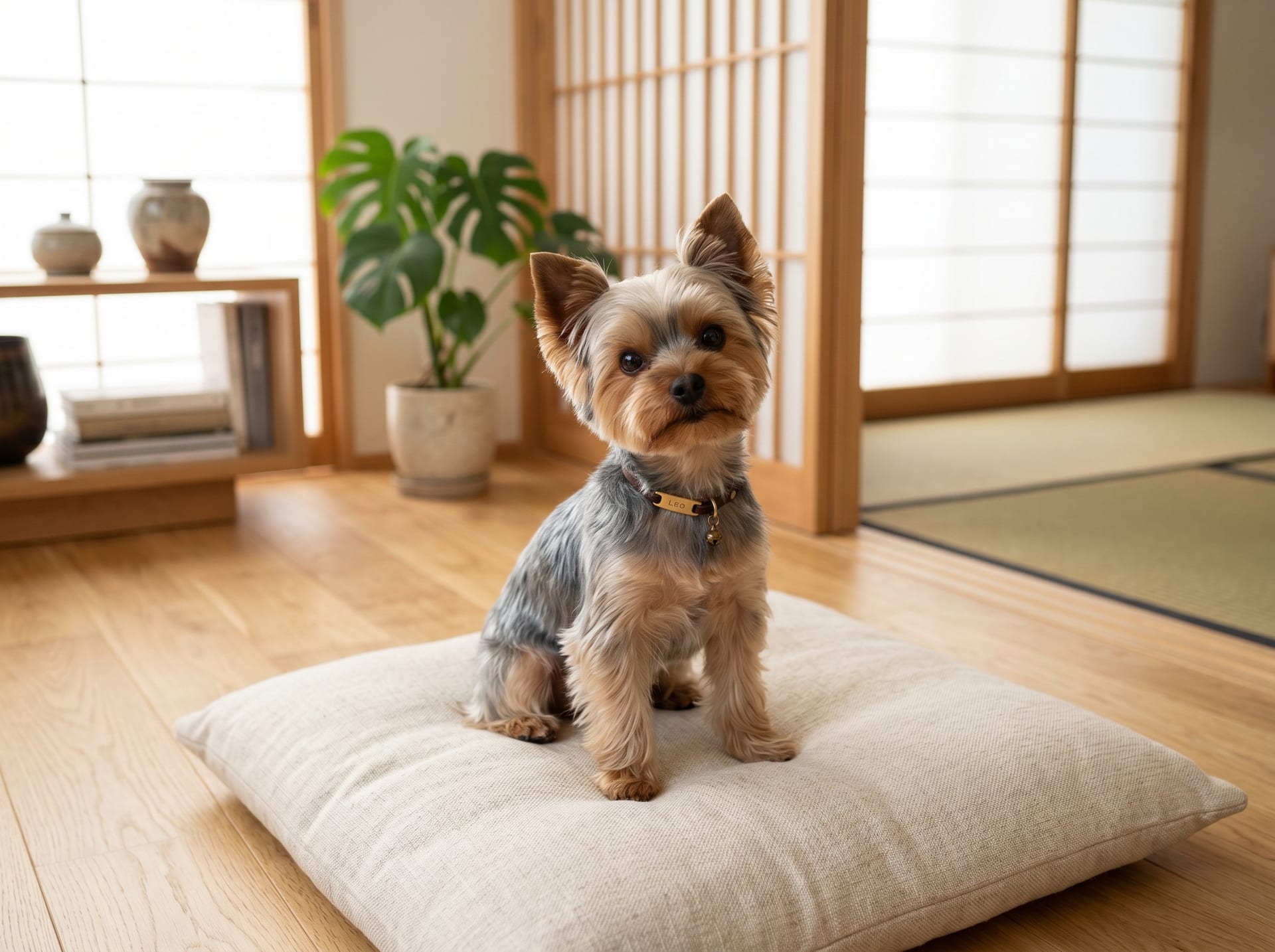
What is Patellar Luxation (Patellar Dislocation)?
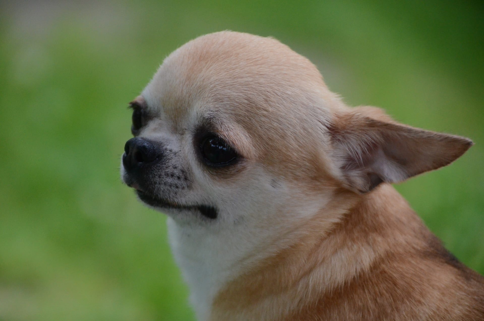
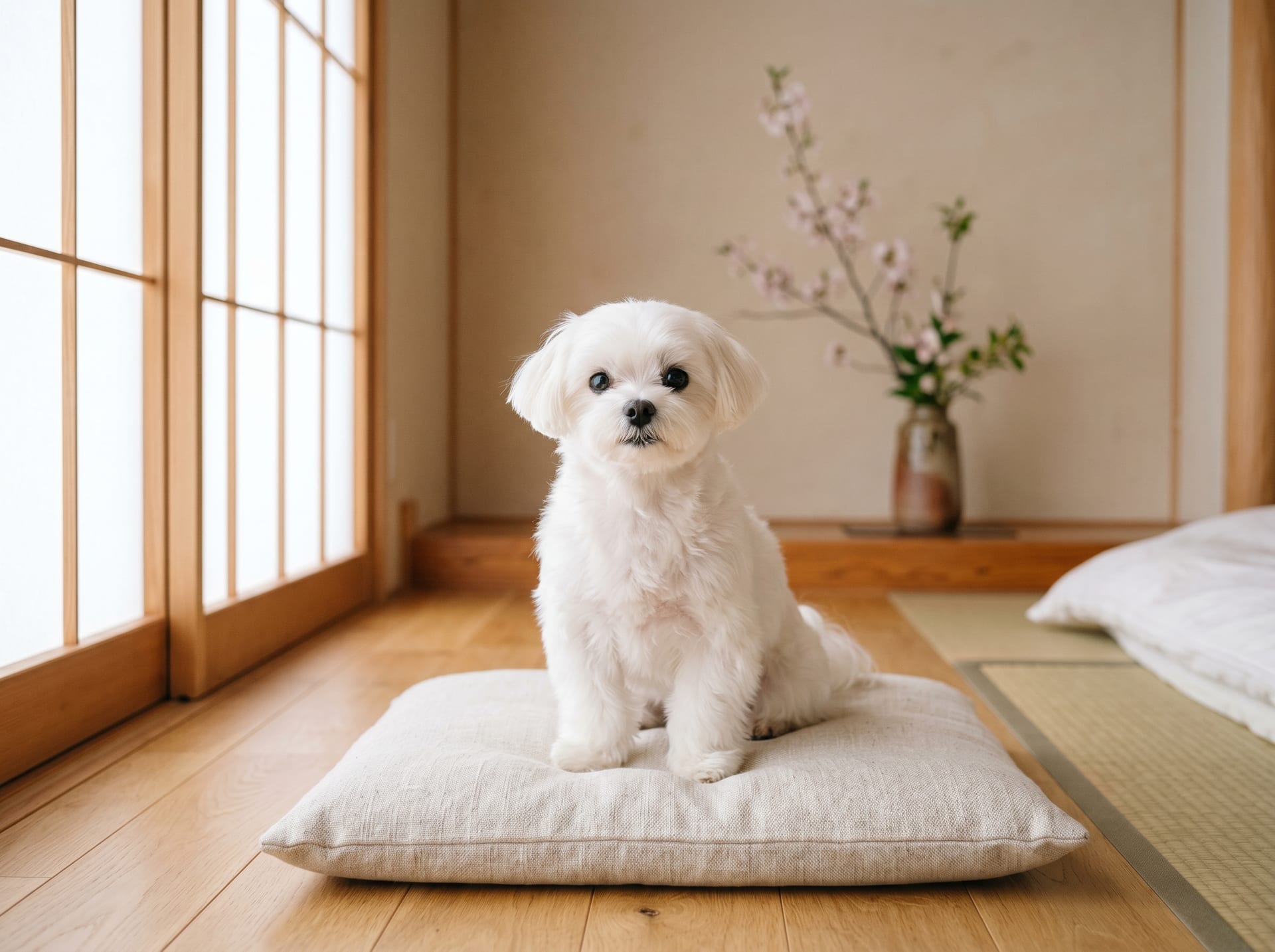
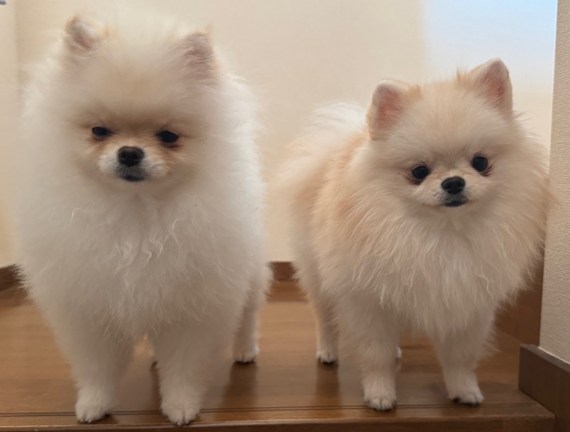
Patellar luxation (Luxating Patella) is an orthopedic condition in which the patella (kneecap), located on the front of the knee, slips medially or laterally from its normal groove. In Japan, it is extremely common in small breed dogs such as Toy Poodles, Chihuahuas, Pomeranians, Yorkshire Terriers, and Maltese dogs, with such a high incidence rate that it is sometimes called "the national disease of small dogs."
The causes are largely due to congenital skeletal structural issues. The condition develops from accumulated skeletal deformities, such as a shallow femoral trochlear groove or misalignment of the tibial tuberosity. In many cases, symptoms first appear within a few months to several years after birth, and may worsen as the dog grows. Acquired factors can also contribute, including obesity, repeated jumping, and trauma from living on hardwood floors.
In mild cases, there is little impact on daily life; however, if left untreated, it can progress to articular cartilage wear, cranial cruciate ligament rupture, and skeletal deformity, eventually leading to difficulty walking. Early detection and early treatment significantly influence the prognosis.
Grading (Severity Classification)
Patellar luxation is classified into four grades (Grade 1–4) based on severity. Diagnosis is made using a combination of palpation and radiographic (X-ray) examination.
Grade 1
The patella remains in its normal position at rest. While it can be displaced with manual pressure, it spontaneously returns to its normal position when pressure is released. There are minimal to no symptoms in daily life, and periodic monitoring is the primary approach.
Grade 2
The patella may spontaneously dislocate during walking or running. A characteristic sign is "skip walking" (lifting one hind leg for 1–2 steps and moving on three legs). While it often spontaneously returns, increased dislocation frequency leads to accumulated strain on muscles and ligaments.
Grade 3
The patella is chronically dislocated. While it can be manually reduced (pushed back), it immediately dislocates again. Lameness (limping) is observed daily, and muscle atrophy begins to develop.
Grade 4
The patella is completely and permanently dislocated and cannot be manually reduced. The skeletal structure itself is deformed, and many dogs develop bowleggedness (varus) or knock-knees (valgus), making normal walking very difficult. At this stage, complete recovery is difficult even with surgery, making early intervention critically important.
Surgical Necessity and Methods
Surgery is considered for dogs with Grade 2 or higher that show impact on daily life, or for young dogs (1–5 years old) with predicted deterioration. Consult a specialist veterinarian early if multiple signs below are observed:
- Lameness occurs repeatedly
- The affected area causes pain when touched (pain response)
- Radiographs confirm progression of arthritis or skeletal deformity
- Obvious muscle atrophy has developed
Primary surgical methods combine multiple techniques. Tibial tuberosity transposition (TTT) corrects bone position so the patella tracks normally; trochlear groove deepening (trochleoplasty) reshapes the groove to provide better patellar stability; and capsular plication tightens periarticular tissues to prevent redislocation.
Costs typically range from ¥150,000 to ¥300,000 per limb and vary depending on the clinic, grade, and surgical techniques used. While bilateral simultaneous surgery is sometimes performed, many veterinarians prefer staggered surgery due to the increased difficulty of post-operative rest management.
Post-operative Rehabilitation and Daily Management
Strict confinement for 2–4 weeks after surgery is the top priority. Excessive activity during this period risks bone slippage.
Basic Post-operative Management
- Continue wearing an Elizabethan collar until suture removal
- Enforce strict crate confinement
- Apply cold compresses to the surgical site several times daily to reduce inflammation (follow veterinarian instructions)
Environmental Modifications
Hardwood floors are a high-risk environment not just post-operatively, but for all dogs with patellar luxation. Install non-slip mats throughout hallways and living areas and prohibit jumping on furniture or over obstacles. Use pet ramps for elevation changes. Since obesity significantly increases joint stress, maintaining appropriate body weight is essential regardless of surgical status.
Rehabilitation Exercises
Gradual rehabilitation begins after suture removal and bone union confirmation (4–8 weeks post-op and beyond). Underwater treadmills or shallow pool walking effectively recover muscle strength while reducing joint loading through buoyancy. On land, begin with short-distance flat ground walking and gradually increase distance and duration on a weekly basis.
Managing Without Surgery: Cases and Methods
Grade 1 or mild Grade 2 cases can often be stabilized long-term with conservative management (non-surgical treatment).
The cornerstone of conservative management is muscle strengthening. Building lower limb muscles, especially the quadriceps, provides stabilizing force to keep the patella in its proper position. Daily flat-ground walks, core training using balance disks or exercise balls, and cavaletti (walking over small raised poles) are all effective approaches.
Glucosamine, chondroitin, and omega-3 fatty acids (fish oil) are sometimes used as supplements to protect articular cartilage. However, scientific evidence varies between individuals, so they should be considered supplementary only.
Regular check-ups every 3–6 months should confirm any grade changes, and treatment plans should be promptly revised if deterioration is detected.
Checklist When Acquiring a Dog from a Breeder
Since patellar luxation has a strong genetic component, verify the following when acquiring a puppy from a breeder:
- Parental patellar screening history: Reputable breeders conduct grade evaluations on breeding dogs
- JKC/OFA breeding standards: Select breeders who exclude Grade 2 or higher dogs from breeding programs
- Veterinary health certificate at 45–60 days old: Confirm orthopedic screening is performed before puppy transfer
- Rearing environment: Ensure excessive hardwood floor exposure or repeated jumping from heights is avoided
While patellar luxation cannot be completely prevented, appropriate breeding practices, early detection, and consistent management can effectively maintain a high quality of life (QOL) for the dog over the long term. Pay close attention to changes in daily gait, and consult a veterinary clinic early if skip walking is observed.